Pleural Mesothelioma Pet Ct - Performance Characteristics Of The Digital Umi550 Pet Ct System According To The Nema Nu2 2018 Standard Ejnmmi Physics Full Text - mesothelioma tests commonly include imaging scans, such as mri and ct scans, and biopsies such as pleural aspiration or thoracoscopy.
Pleural Mesothelioma Pet Ct - Performance Characteristics Of The Digital Umi550 Pet Ct System According To The Nema Nu2 2018 Standard Ejnmmi Physics Full Text - mesothelioma tests commonly include imaging scans, such as mri and ct scans, and biopsies such as pleural aspiration or thoracoscopy.. Pehlivan b, topkan e, onal c, et al. This lets the doctor compare areas of higher radioactivity on the pet. pleural mesothelioma can cause fluid to build up around the lungs in the chest (called a pleural effusion). Your gp will conduct a physical examination and order tests. In this prospective study, pet results in patients with pleural abnormalities on ct were compared with histologic results.
In some cases, this leads to pleural mesothelioma. Fdg, pet/ct, pleura, pleural metastases, mesothelioma, pleurodesis. A pet scan is ideal for use as a diagnostic device for pleural as well as abdominal mesothelioma. Malignant peritoneal mesothelioma is an uncommon primary tumor of the peritoneal lining. Mri exams complement ct scanning in some patients, providing better delineation of soft tissues and allowing imaging in the sagittal and coronal planes.
Jcm Free Full Text Impact Of Pet Ct For Assessing Response To Immunotherapy A Clinical Perspective Html from www.mdpi.com A short summary of this paper. Feigen m, lee st, lawford c, et al. Symptoms include chest pain, coughing, and shortness of breath. Patients are injected with a radioactive tracer isotope combined with some form of glucose. • mesothelioma may be confused with metastatic adenocarcinoma, other mets to pleura, such as thymoma metastatic to pleura • mesothelioma: Malignant mesothelioma (mm) is an uncommon neoplasm with a poor prognosis usually associated with asbestos exposure. pet has proven to be superior as a diagnostic tool in several malignancies. There is a global epidemic of malignant pleural mesothelioma underway, and incidence rates are predicted to peak in the next few years.
A cough that never gets better or pain in the chest wall with no obvious cause.
Malignant pleural mesothelioma is an aggressive malignancy of the pleural surface, predominantly caused by prior asbestos exposure. In cases of peritoneal mesothelioma, fluid can build up in the abdomen (called ascites). Current perspectives maria bonomi,1 costantino de filippis,2 egesta lopci,3 letizia gianoncelli,1 giovanna rizzardi,4 eleonora cerchiaro,1 luigi bortolotti,4 alessandro zanello,2 giovanni luca ceresoli1 1department of oncology, thoracic and gu oncology unit, 2department of radiology, cliniche humanitas gavazzeni, bergamo, 3nuclear medicine. Fdg, pet/ct, pleura, pleural metastases, mesothelioma, pleurodesis. Symptoms include chest pain, coughing, and shortness of breath. Eligible patients had pleural thickening on ct and were medically fit for surgical diagnostic procedures. Clinical staging of malignant pleural mesothelioma: This condition is usually associated with occupational exposure to asbestos.wagner et al connected asbestos to mesothelioma in a classic 1960 study of 33 patients with mesothelioma who were exposed to asbestos in a mining area in south africa's north western cape province. Malignant pleural mesothelioma is a dangerous cancer that affects the lining of the lungs (pleura). A pet scan is the best method to determine if mesothelioma has spread to other parts of the body. Mri exams complement ct scanning in some patients, providing better delineation of soft tissues and allowing imaging in the sagittal and coronal planes. Patients received 2.5 ml fluorescein intravenously preceding the procedure. Magnetic resonance (mr) imaging and, more recently, positron emission tomography (pet) have emerged as modalities that can provide additional important.
A short summary of this paper. In cases of peritoneal mesothelioma, fluid can build up in the abdomen (called ascites). mesothelioma tests commonly include imaging scans, such as mri and ct scans, and biopsies such as pleural aspiration or thoracoscopy. Current perspectives maria bonomi,1 costantino de filippis,2 egesta lopci,3 letizia gianoncelli,1 giovanna rizzardi,4 eleonora cerchiaro,1 luigi bortolotti,4 alessandro zanello,2 giovanni luca ceresoli1 1department of oncology, thoracic and gu oncology unit, 2department of radiology, cliniche humanitas gavazzeni, bergamo, 3nuclear medicine. Fdg, pet/ct, pleura, pleural metastases, mesothelioma, pleurodesis.
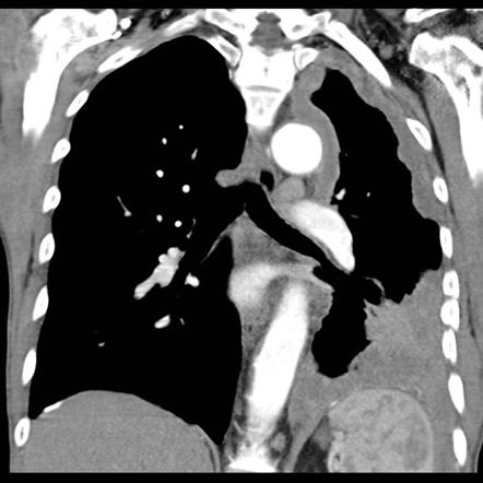
Management Of Pleural Mesothelioma Thoracic Key from i2.wp.com Introduction talc pleurodesis (tp) is a procedure first described by bethune 1 in 1935 as a means to anchor the lung during lobectomy 2 . mesothelioma usually starts in the pleura of the lungs, but it can also start in the abdomen or other organs. Early detection of the fatal and incurable mesothelioma and the later provision of radiation, surgical and palliative asbestosis treatments are known to help a patient to have the best possible chance to extend and enhance the quality of life remaining. Malignant peritoneal mesothelioma is an uncommon primary tumor of the peritoneal lining. A chest ct was performed afterwards, which demonstrated a small, homogeneous and smooth mass arising from the pleural surface (figure 2). Positron emission tomography (pet) scan: mesothelioma has a long latency period of 20 to 40 years, and many patients do not have symptoms until the disease is in its later stages, when metastasis is more likely to occur. Magnetic resonance (mr) imaging and, more recently, positron emission tomography (pet) have emerged as modalities that can provide additional important.
Introduction talc pleurodesis (tp) is a procedure first described by bethune 1 in 1935 as a means to anchor the lung during lobectomy 2 .
Some machines can do both a pet and ct scan at the same time. Malignant pleural mesothelioma is a dangerous cancer that affects the lining of the lungs (pleura). Doctors can make a metastatic mesothelioma diagnosis through radiology imaging scans such as mri, ct or pet. The main symptoms are shortness of breath, pain when breathing, chest/shoulder/upper arm pain, loss of appetite, weight loss, and persistent cough or bouts of pneumonia. Other abdominal subtypes (also discussed separately) include: Because mesothelioma may spread to the diaphragm, an mri may be used to look at the diaphragm, the muscle used for breathing, which separates the chest from the abdomen. The group made the recommendation that one must exercise caution when interpreting surveillance images from patients with talc pleurodesis in the setting of mesothelioma as. Patients received 2.5 ml fluorescein intravenously preceding the procedure. The visceral pleura covers each lung surface, and the parietal pleura covers the inner surface of the thoracic cavity. The purpose was to assess the accuracy of ct scan based preoperatively measured tumor volume and thickness compared to actual tumor weight of resected mpm specimen and pathologically assessed tumor. There is a global epidemic of malignant pleural mesothelioma underway, and incidence rates are predicted to peak in the next few years. Imaging plays an essential role in the evaluation of malignant pleural mesothelioma (mpm). This retrospective study aimed to investigate the prognostic value of the suvmax in patients with mpm.
Doctors can even use it to find out if a tumor has spread to lymph nodes that are far from where the mesothelioma first appeared. • mesothelioma may be confused with metastatic adenocarcinoma, other mets to pleura, such as thymoma metastatic to pleura • mesothelioma: The visceral pleura covers each lung surface, and the parietal pleura covers the inner surface of the thoracic cavity. Imaging plays an essential role in the evaluation of malignant pleural mesothelioma (mpm). Early detection of the fatal and incurable mesothelioma and the later provision of radiation, surgical and palliative asbestosis treatments are known to help a patient to have the best possible chance to extend and enhance the quality of life remaining.
Mesothelioma Radiology Reference Article Radiopaedia Org from prod-images-static.radiopaedia.org And patients should undergo pleural morphology and immunohistochemistry as soon as possible, which are helpful for timely diagnosis. Computed tomography is the primary imaging modality used for the diagnosis and staging of mpm. The emphasis will be on pet/ct. The group made the recommendation that one must exercise caution when interpreting surveillance images from patients with talc pleurodesis in the setting of mesothelioma as. Patients are injected with a radioactive tracer isotope combined with some form of glucose. The main symptoms are shortness of breath, pain when breathing, chest/shoulder/upper arm pain, loss of appetite, weight loss, and persistent cough or bouts of pneumonia. This condition is usually associated with occupational exposure to asbestos.wagner et al connected asbestos to mesothelioma in a classic 1960 study of 33 patients with mesothelioma who were exposed to asbestos in a mining area in south africa's north western cape province. In some cases, this leads to pleural mesothelioma.
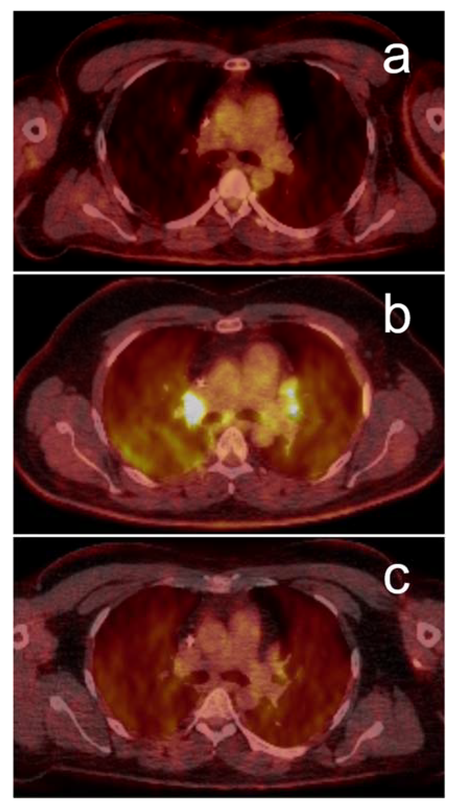
One of the best and most widely used scans for detecting and diagnosing mesothelioma and other kinds of cancer is the positron emission tomography scan, known as a pet scan.
This retrospective study aimed to investigate the prognostic value of the suvmax in patients with mpm. pleural mesothelioma is caused by inhaling asbestos fibers that get lodged into the protective lining of the lungs (the pleura) which cause genetic mutations in the surrounding cells. The pleura are the pair of membranous linings surrounding the lungs. These areas are highlighted in order to produce a contrast to the patient's normal, healthy tissue. This is due to the details provided in the images of the peritoneum (the lining of the abdomen) and the pleura (the lining of the lung). Other abdominal subtypes (also discussed separately) include: Early detection of the fatal and incurable mesothelioma and the later provision of radiation, surgical and palliative asbestosis treatments are known to help a patient to have the best possible chance to extend and enhance the quality of life remaining. One of the best and most widely used scans for detecting and diagnosing mesothelioma and other kinds of cancer is the positron emission tomography scan, known as a pet scan. This article summarises the epidemiology and pathogenesis of malignant pleural mesothelioma, before describing some key factors. The purpose was to assess the accuracy of ct scan based preoperatively measured tumor volume and thickness compared to actual tumor weight of resected mpm specimen and pathologically assessed tumor. These fibers get lodged into the protective lining of the lungs (the pleura), causing genetic mutations in the surrounding cells. Eligible patients had pleural thickening on ct and were medically fit for surgical diagnostic procedures. • understaging common with pet/ct but pet useful to determine


0 Comments